KRT4 Recombinant Monoclonal Antibody
-
中文名稱:KRT4 Recombinant Monoclonal Antibody
-
貨號:CSB-RA909139A0HU
-
規格:¥1320
-
圖片:
-
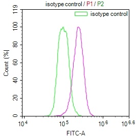
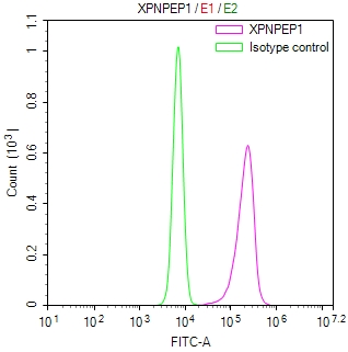
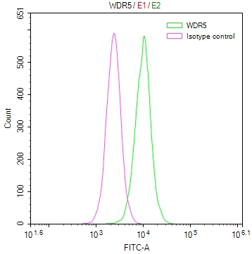
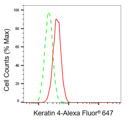 Flow cytometric analysis of keratin 4 expression in H9C2 cells using keratin 4 antibody. Green, isotype control; red, keratin 4.
 -
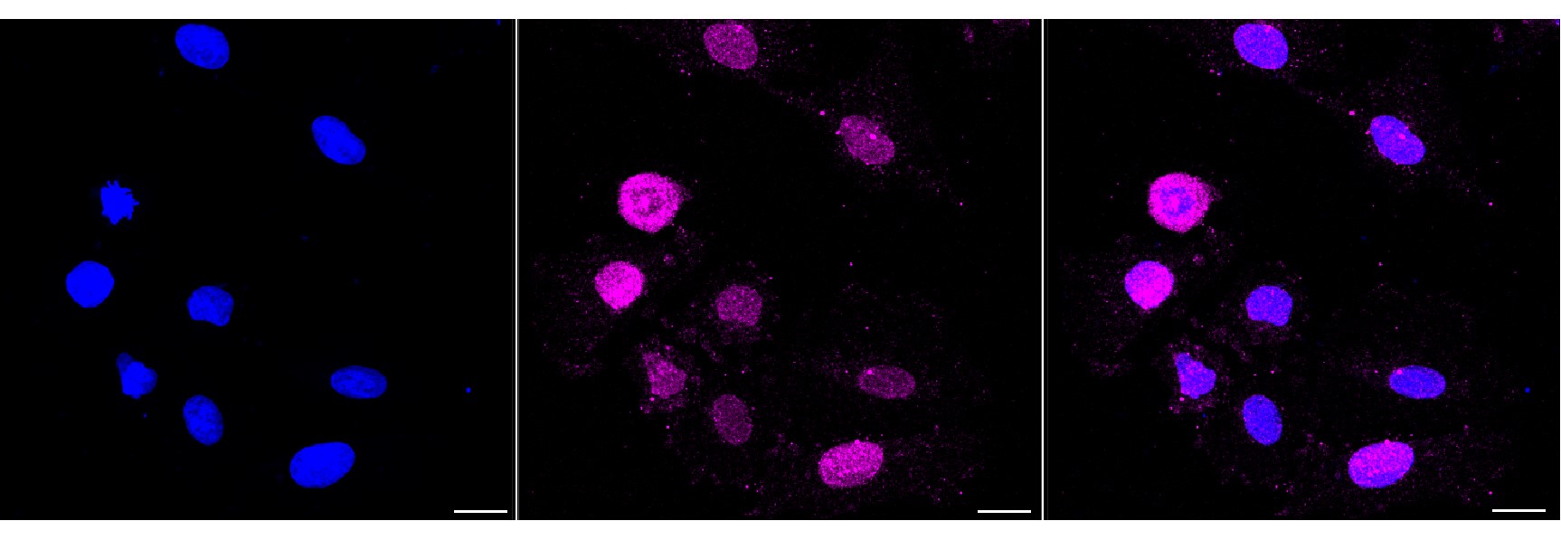 Immunocytochemical staining of H9c2 cells with Keratin 4 antibody. Nuclei were stained blue with DAPI; Keratin 4 was stained magenta with Alexa Fluor? 647. Images were taken using Leica stellaris 5. Protein abundance based on laser Intensity and smart gain: Medium. Scale bar, 20 μm.
 -
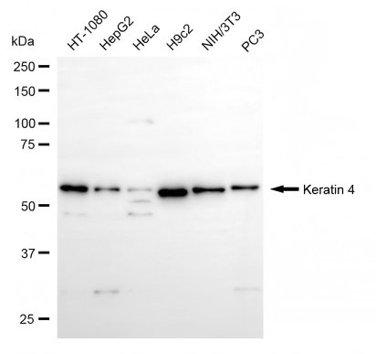 Western blotting analysis using keratin 4 antibody. Total cell lysates (30 μg) from various cell lines were loaded and separated by SDS-PAGE. The blot was incubated with keratin 4 antibody and HRP-conjugated goat anti-rabbit secondary antibody respectively.
-
-
其他:
產品詳情
-
Uniprot No.:
-
基因名:KRT4
-
別名:KRT4; Keratin 4; CYK4; K4; Keratin, Type II Cytoskeletal 4; CK4; Type-II Keratin Kb4; Cytokeratin 4; CK-4; Keratin 4, Type II; Cytokeratin-4; Keratin-4; WSN1
-
反應種屬:Human, Mouse, Rat
-
免疫原:Recombinant Human KRT4 protein
-
免疫原種屬:Homo sapiens (Human)
-
標記方式:Non-conjugated
-
克隆類型:Monoclonal
-
抗體亞型:Rabbit IgG
-
純化方式:Affinity-chromatography
-
克隆號:7E10
-
濃度:It differs from different batches. Please contact us to confirm it.
-
保存緩沖液:Rabbit IgG in PBS (pH 7.4) containing 50% glycerol, and 0.02% sodium azide.
-
產品提供形式:Liquid
-
應用范圍:ELISA, WB, FC, ICC
-
推薦稀釋比:
Application Recommended Dilution WB 1:1000-1:5000 FC 1:200-1:2000 ICC 1:100-1:1000 -
Protocols:
-
儲存條件:Upon receipt, store at -20°C or -80°C. Avoid repeated freeze.
-
貨期:Basically, we can dispatch the products out in 1-3 working days after receiving your orders. Delivery time maybe differs from different purchasing way or location, please kindly consult your local distributors for specific delivery time.
-
用途:For Research Use Only. Not for use in diagnostic or therapeutic procedures.
產品評價

相關產品
靶點詳情
-
相關疾病:White sponge nevus 1 (WSN1)
-
蛋白家族:Intermediate filament family
-
組織特異性:Detected in the suprabasal layer of the stratified epithelium of the esophagus, exocervix, vagina, mouth and lingual mucosa, and in cells and cell clusters in the mucosa and serous gland ducts of the esophageal submucosa (at protein level). Expressed wide
-
數據庫鏈接:
Most popular with customers
-
YWHAB Recombinant Monoclonal Antibody
Applications: ELISA, WB, IHC, IF, FC
Species Reactivity: Human, Mouse, Rat
-
Phospho-YAP1 (S127) Recombinant Monoclonal Antibody
Applications: ELISA, WB, IHC
Species Reactivity: Human
-
-
-
-
-
VDAC1 Recombinant Monoclonal Antibody
Applications: ELISA, WB, IHC
Species Reactivity: Human, Mouse, Rat
-

VCP Recombinant Monoclonal Antibody
Applications: ELISA, WB, IHC, IF, IP
Species Reactivity: Human, Rat